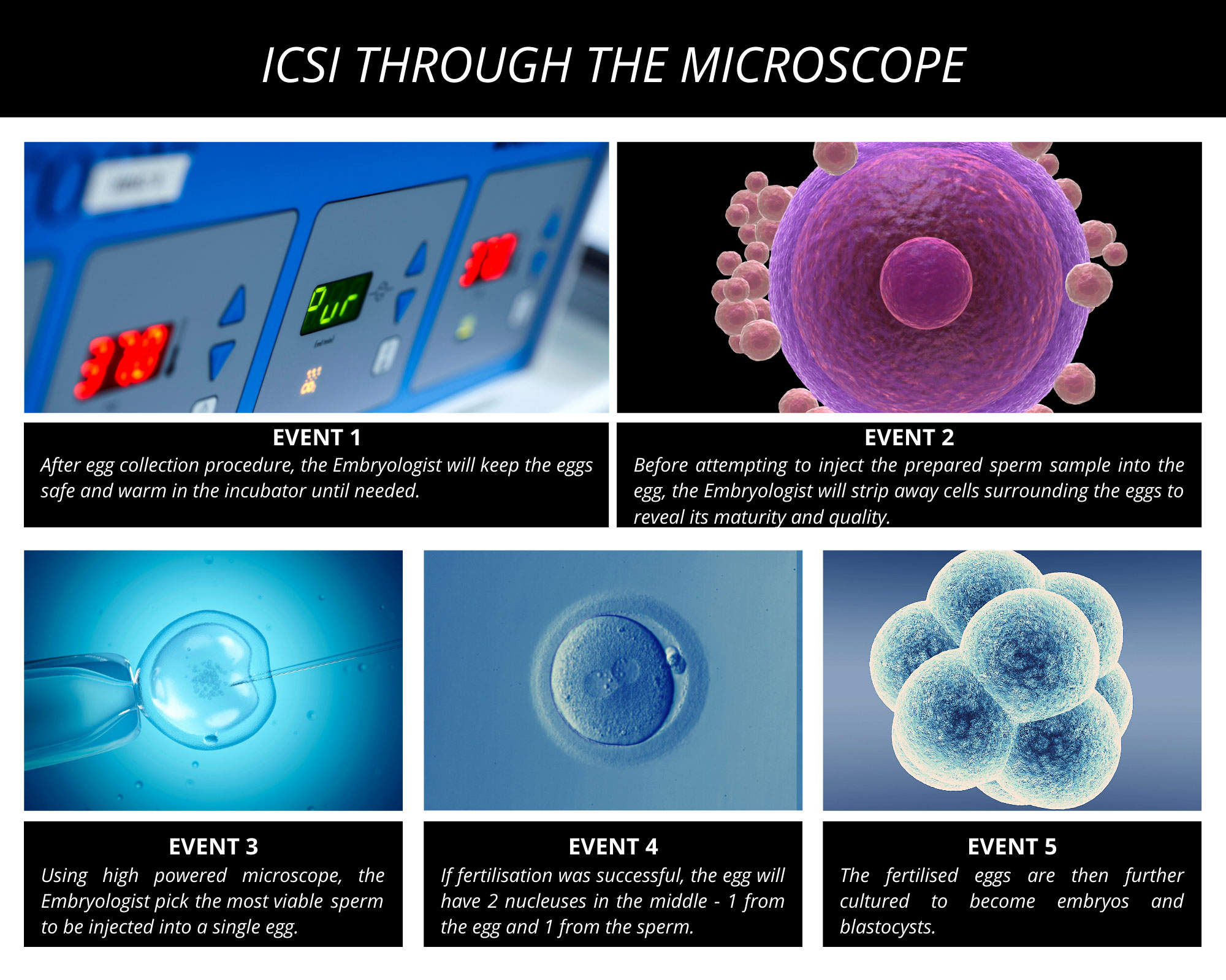
The illustration above explains the process of ICSI (Intracytoplasmic Sperm Injection) as it is performed. We use ICSI when all other measures have failed to benefit the male partner who has poor sperm.
In ICSI, a single sperm is injected into the woman’s egg to cause it to fertilise and become an embryo. The first part of the ICSI technique is to immobilize the egg. The pipette (fine glass tube) on the left can be seen holding the egg by suction.
The ICSI injector on the right then picks up a single sperm and slowly penetrates the thick wall of the egg and deposits the sperm within it.
Finally the egg is released and will then spend a further three to five days inside an incubator which mimics the woman’s body. Here it will slowly develop into an embryo.
Once the ICSI injection is done, there is no difference in the development of the embryo when compared to IVF. The pregnancy rate with ICSI is also similar to that with IVF.
The whole ICSI process is performed under a high powered microscope so what you see in the video above is magnified 400 times.